Enabling Functional Analysis of Cellular Immunology
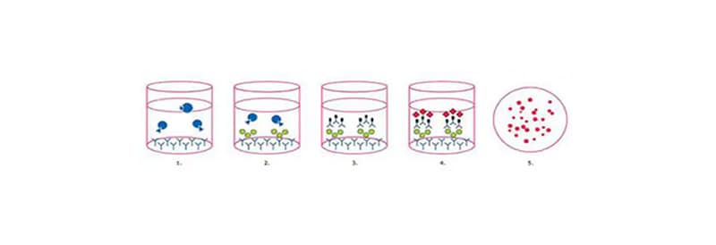
Under optimal conditions, the enzyme-linked immunosorbent spot (ELISpot) assay enables visualization of multiple secretory products from a single responding cell. Thus, the ELISpot provides both qualitative (type of immune protein) and quantitative (number of responding cells) information. By virtue of this assay’s unsurpassed sensitivity, frequency analysis of rare cell populations (e.g., antigen-specific responses) which was not possible before is now relatively easy. Recent improvements to the design of multiwell microplates, including use of membranes with reduced background fluorescence, have bolstered the widespread application of EliSpot assays.
When assay sensitivity, ease of use, and cost are all taken into consideration, the EliSpot platform is likely the superior choice for the development of multifunctional T-cell assays for the research, therapeutic, and diagnostic communities.
The popularity of this assay has seen a resurgence in recent years as researchers attempt to gain a better understanding of immune responses in a variety of applications including study of immunological memory and vaccine development.
ELISpot Optimization
While ELISpot assays permit frequency determination for very rare events, data interpretation can become ambiguous when spot numbers in antigen-containing wells are low, spot counts in negative control wells are elevated, and particularly when both occur simultaneously. Thus, the primary task, even before statistics are employed, must be the optimization of basic assay parameters and reagents to maximize the signal-to-noise ratio. While the use of highly specific ELISpot–validated Ab pairs is key to assay success, proper consideration and execution of a number of other steps are required to ensure optimal performance.
Choice of Plate (Membrane)
PVDF membrane plates (Merck Catalog Nos. MSIPS4W10, MSIPS4510, MAIPSWU10, MAIPS4510) are recommended, over a mixed cellulose ester format (Merck Catalog No. MSHAS4510), due to slightly improved binding of capture Ab and superior performance in spot detection, particularly for fluorescent applications. The one drawback of PVDF plates is the extreme hydrophobicity of the material, a property that may necessitate pre-wetting with alcohol prior to addition of the coating Ab. Since the mixed cellulose membrane is hydrophilic, ELISpot assays can be performed without pre-wetting.
Negative/Positive Controls
Relevant controls are crucial to measuring Ag-specific responses via ELISpot. Negative controls routinely consist of cells cultured without stimuli, whereas polyclonal T-cell activators are commonly used as positive controls to confirm both cell and assay functionality. Positive controls include anti-CD3/CD28 Abs, phytohemagglutinin (PHA), and concanavalin A (ConA). These activators induce secretion of many common cytokines including IFNγ, IL-2 (Th1), IL-4, IL-5, IL-10, and IL-13 (Th2).
Another common control is the commercially available CEF (Cytomegalovirus, Epstein-Barr virus, Influenza virus) peptide pool. These consist of multiple epitopes from each of the three viruses, to which most healthy individuals (~ 90%) possess CD8-responding T-cells.
As we can only outline some aspects in this article, we would like to refer you to our ELISpot White Paper where the assay protocol will be discussed in detail with an emphasis on standard best practice and troubleshooting.
Related Products

Catalog Number 10492352

Catalog Number 10169794