missing translation for 'onlineSavingsMsg'
Learn More
Learn More

p53 Antibody (BP53-12), Novus Biologicals™
Mouse Monoclonal Antibody has been used in 3 publications
307.00 € - 561.00 €
Specifications
| Antigen | p53 |
|---|---|
| Clone | BP53-12 |
| Concentration | 0.2 mg/ml |
| Dilution | Western Blot 0.5-1ug/ml, Simple Western 10 ug/ml, Flow Cytometry 0.5-1ug/million cells in 0.1ml, Immunocytochemistry/Immunofluorescence 1-2ug/ml, Immunoprecipitation 1-2ug/500ug protein lysate, Immunohistochemistry-Paraffin 0.5-1.0ug/ml, Immunohistochemistry-Frozen 0.5-1.0ug/ml |
| Applications | Western Blot, Flow Cytometry, Immunocytochemistry, Immunofluorescence, Immunoprecipitation |
| Product Code | Brand | Quantity | Price | Quantity & Availability | |||||
|---|---|---|---|---|---|---|---|---|---|
| Product Code | Brand | Quantity | Price | Quantity & Availability | |||||
|
18465861
|
Novus Biologicals
NBP2-29453-20ug |
20 ug |
307.00 €
20µg |
Please sign in to purchase this item. Need a web account? Register with us today! | |||||
|
18785193
|
Novus Biologicals
NBP2-29453 |
561.00 €
0.10mg |
Please sign in to purchase this item. Need a web account? Register with us today! | ||||||
Description
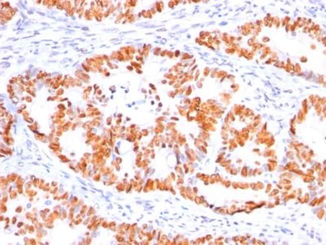
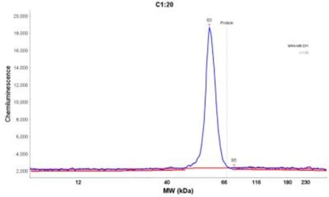
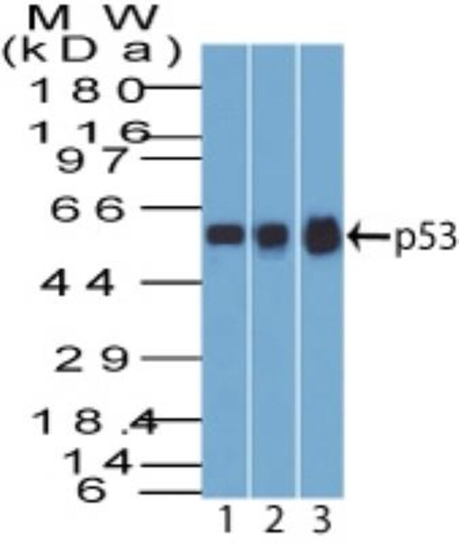
p53 Monoclonal specifically detects p53 in Human, Canine, Chicken, Hamster, Monkey, Mouse (Negative), Rat (Negative) samples. It is validated for Western Blot, Simple Western, Immunohistochemistry, Immunohistochemistry-Paraffin.Specifications
| p53 | |
| 0.2 mg/ml | |
| Western Blot, Flow Cytometry, Immunocytochemistry, Immunofluorescence, Immunoprecipitation | |
| Unconjugated | |
| Mouse | |
| Apoptosis, Cancer, Cell Cycle and Replication, Cellular Markers, Checkpoint signaling, Core ESC Like Genes, DNA Double Strand Break Repair, DNA Repair, HIF Target Genes, Hypoxia, Neuroscience, Neurotransmission, p53 Pathway, Phospho Specific, Stem Cell Markers, Transcription Factors and Regulators, Tumor Suppressors | |
| P04637 | |
| 7157 | |
| Recombinant human wild-type p53 protein (Uniprot: P04637) | |
| Primary | |
| Store at 4C. | |
| 53 kDa |
| BP53-12 | |
| Western Blot 0.5-1ug/ml, Simple Western 10 ug/ml, Flow Cytometry 0.5-1ug/million cells in 0.1ml, Immunocytochemistry/Immunofluorescence 1-2ug/ml, Immunoprecipitation 1-2ug/500ug protein lysate, Immunohistochemistry-Paraffin 0.5-1.0ug/ml, Immunohistochemistry-Frozen 0.5-1.0ug/ml | |
| Monoclonal | |
| Purified | |
| RUO | |
| Human, Mouse, Canine, Chicken, Hamster, Monkey, Mouse (Negative), Rat (Negative) | |
| Antigen NY-CO-13, FLJ92943, LFS1TRP53, p53, p53 tumor suppressor, P53cellular tumor antigen p53, Phosphoprotein p53, transformation-related protein 53, tumor protein p53, Tumor suppressor p53 | |
| TP53 | |
| IgG2a κ | |
| Protein A or G purified | |
| This monoclonal antibody reacts with an N-terminal epitope (aa 16-25) of both wild type and mutated p53. Mutation and/or allelic loss of p53 is one of the causes of a variety of mesenchymal and epithelial tumors. If it occurs in the germ line, such tumors run in families. In most transformed and tumor cells the concentration of p53 is increased 51000 fold over the minute concentrations (1000 molecules cell) in normal cells, principally due to the increased half-life (4 h) compared to that of the wild-type (20 min). p53 Localizes in the nucleus, but is detectable at the plasma membrane during mitosis and when certain mutations modulate cytoplasmic/nuclear distribution. Mutations arise with an average frequency of 70% but incidence varies from zero in carcinoid lung tumors to 97% in primary melanomas. High concentrations of p53 protein are transiently expressed in human epidermis and superficial dermal fibroblasts following mild ultraviolet irradiation. Positive nuclear staining with p53 antibody has been reported to be a negative prognostic factor in breast carcinoma, lung carcinoma, colorectal, and urothelial carcinoma. Anti-p53 positivity has also been used to differentiate uterine serous carcinoma from endometrioid carcinoma as well as to detect intratubular germ cell neoplasia. |
For Research Use Only
Spot an opportunity for improvement?Share a Content Correction
Product Content Correction
Your input is important to us. Please complete this form to provide feedback related to the content on this product.
Product Title